Cochin Cardiac Club

Health Blog by Dr.Uday Nair

COR PULMONALE

Right-sided heart failure


Cor pulmonale is failure of the right side of the heart brought on by long-term high blood pressure in the pulmonary arteries and right ventricle of the heart.


Causes

Normally, the left side of the heart produces a higher blood pressure in order to pump blood to the body. The right side of the heart pumps blood through the lungs under much lower pressure.
Any condition that leads to prolonged high blood pressure in the arteries of the lungs (called pulmonary hypertension) puts a strain on the right side of the heart. When the right ventricle is unable to properly pump against these abnormally high pressures, it is called cor pulmonale.
Almost any chronic lung disease or condition causing prolonged low blood oxygen levels can lead to cor pulmonale. A few of these causes include:
  • Central sleep apnea
  • Chronic obstructive pulmonary disease (COPD)
  • Chronic thromboembolic pulmonary disease
  • Cystic fibrosis
  • Interstitial lung disease
  • Kyphoscoliosis
  • Obstructive sleep apnea
  • Pneumoconiosis
  • Primary pulmonary hypertension
  • Pulmonary vascular disease
  • Secondary pulmonary hypertension


    Symptoms


    • Chest discomfort, usually in the front of the chest
    • Exercise intolerance
    • Shortness of breath
    • Swelling of the feet or ankles
    • Symptoms of underlying disorders (wheezing, coughing)

Signs and tests


  • Abnormal fluid collection in the abdomen
  • Abnormal heart sounds
  • Bluish color to the skin (cyanosis)
  • Enlargement of the liver
  • Swelling (distension) of the neck veins, indicating high right-heart pressures
  • Swelling of the ankles
The following tests may help diagnose cor pulmonale:
  • Blood antibody tests
  • Blood test for brain natriuretic peptide (BNP)
  • Chest x-ray
  • CT scan of the chest
  • Echocardiogram
  • Lung biopsy (rarely performed)
  • Measurement of blood oxygen by arterial blood gas (ABG)
  • Pulmonary function tests
  • Right heart catheterization
  • Ventilation and perfusion scan of the lungs (V/Q scan)
Treatment


Treatment is directed at the illness that is causing cor pulmonale. Supplemental oxygen may be prescribed to increase the level of oxygen in the blood.
There are many medicines available to treat cor pulmonale.
  • Bosentan or sildenafil may be given by mouth
  • Calcium channel blockers are often used to treat early cases
  • Prostacyclin may be given through injection or breathing in (inhalation)
Blood thinning (anticoagulant) medications may also be prescribed. Surgery may be needed to reverse heart defects that cause the condition. In very advanced cases, a heart and lung transplant may be advised.


Expectations (prognosis)


The outcome depends on the cause of the condition. Giving oxygen often improves symptoms, stamina, and survival.
Treating primary pulmonary hypertension often leads to greater stamina and a longer life. In some cases, a lung transplant or heart-lung transplant can extend survival.


Complications

Progressive pulmonary hypertension and cor pulmonale may lead to:
  • Life-threatening shortness of breath
  • Severe fluid retention
  • Shock
  • Death


Prevention


Avoiding behaviors that lead to chronic lung disease (especially cigarette smoking) may prevent the eventual development of cor pulmonale. Careful evaluation of childhood heart murmurs may prevent cor pulmonale caused by certain heart defects.

Please Note:

Call your doctor if you experience shortness of breath or chest pain.


HEART,KIDNEYS AND UTI


HEART,KIDNEYS AND URINARY TRACT INFECTION.


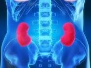
Heart disease is a frequent problem for people with kidney disease. Although heart disease has decreased in  over the past 30 years, patients with chronic kidney disease have not enjoyed the same reduction in the chances of having a heart attack or developing heart failure. People who have diabetes, high blood pressure, high cholesterol, are overweight, smoke, have had a previous heart attack or who have close family members with heart trouble are at the greatest risk of developing heart disease.

Hardening of the arteries is the most frequent cause of heart disease. This process starts with fat deposits in the arteries and may begin at a young age, even in people without kidney disease. Over time, the fat deposits contain calcium and form plaques that can block small arteries, like those in the heart, the brain and the kidneys. Many factors contribute to hardening of the arteries. This article will consider some of the risk factors for the development of hardening of the arteries and heart disease in people with chronic kidney disease and present specific strategies for prevention.

High blood pressure is a well-known risk factor for heart disease and strokes. Blood pressure is usually reported as two numbers, the top number is the systolic blood pressure and the bottom number is the diastolic blood pressure. Both systolic and diastolic blood pressures increase the risk when they are too high, particularly in people who smoke, have diabetes or abnormal cholesterol.
A blood pressure of 130/85 is normal and it is recommended for patients younger than 65. Achieving a normal blood pressure should be the goal of therapy. For people over the age of 65, the risk of strokes is even higher than the risk of a heart attack and a blood pressure of 140/90 is suggested. Patients, who have protein in their urine or decreased kidney function, may benefit from an even lower blood pressure of 125/75, which is thought to slow down kidney damage in people who already have kidney disease.

There are several ways to lower blood pressure without taking medications. A low salt diet, quitting smoking, losing weight, exercising and decreasing the amount of alcohol you drink to no more than one glass of beer, wine, or mixed drink each day. However, most people have to take medicines to get their blood pressure low enough to decrease the risk of heart disease and protect their kidneys. On average, many people with high blood pressure will have to take three or four medications to get their blood pressure under control.

Fat in the blood stream, called lipids, contributes to hardening of the arteries. Cholesterol and triglycerides are fats in the blood stream. Both of these fats are necessary and help the body’s cells to work normally and they provide fuel for the body. Only if the levels get too high, can they contribute to hardening of the arteries. Doctors frequently measure the total cholesterol as an indication of a patient’s risk of developing heart disease. The total cholesterol consists of LDL cholesterol, the so-called bad cholesterol, and HDL, the good cholesterol. Depending on how many other risk factors for heart disease a person has, the target for total cholesterol may be as low as 200 mg/dl. The same level is desirable for triglycerides in people with several risk factors for heart disease.

Lowering the total cholesterol and particularly, the bad cholesterol decreases the risk of heart disease. People with high cholesterol levels should consider changing their diet to avoid fatty foods and other foods high in cholesterol, like red meat. However, diet alone can only lower the cholesterol by a relatively small amount and many people with high cholesterol will need to be on specific medications to lower their cholesterol levels.

Medications that lower cholesterol decrease the risk of heart disease and heart attacks. Although these medications are very effective, they may have side effects that doctors need to monitor. They can also interact with other medications that people with kidney disease may be taking. Consequently, doctors follow blood tests closely in patients taking cholesterol-lowering drugs.

Triglycerides are also a kind of fat normally found in the blood stream. When triglyceride levels are abnormally increased, they may be a risk factor for hardening of the arteries. Decreasing the amount of sugar in the diet may lower triglyceride levels and there are medications, which are also effective for that purpose.

Smoking is a very dangerous risk factor for hardening of the arteries. Smoking decreases the good cholesterol, increases blood pressure, decreases the oxygen in the blood stream and makes it easier for blood to clot in the tiny blood vessels in the heart and brain. Smoking can cause heart attacks and sudden death, particularly in patients who already have heart disease. Smoking may also worsen kidney disease. Quitting smoking is an important way to decrease the risk of strokes and heart attacks. Even among persons who have smoked heavily in the past, the risk of a heart attack can be cut in half regardless of how long or how much they have previously smoked.

Diabetes mellitus or sugar diabetes is an important cause of hardening of the arteries. Diabetes is a leading cause of blockage of the tiny blood vessels in the kidneys, heart, brain and eyes. Diabetes is also the most common cause of kidney failure. Consequently, high blood sugar levels are associated with worsening kidney function, heart attacks, strokes and blindness. Diabetes in adults is often associated with being overweight. Weight loss through sensible diet and exercise decreases the risk of heart disease and may improve diabetes control. In order to achieve the best control of blood sugar, diabetics may take medications by mouth or insulin injections. Some diabetic medicines are not good for patients with kidney disease, because they can cause acid to accumulate in the blood stream. Doctors familiar with kidney patients will avoid using these medicines.

Homocysteine is a chemical normally found in the blood that is important in making proteins. Levels sometime increase in people with chronic kidney disease and may cause hardening of the arteries. Homocysteine blood levels decrease when patients take large doses of folic acid and other B-vitamins. Doctors do not know for sure whether lowering homocysteine levels in this way decreases the risks of heart attacks and strokes.

Individuals with kidney disease are at a high risk of developing hardening of the arteries resulting in heart attacks, heart failure and strokes. Multiple factors are involved. The table below shows multiple strategies to prevent hardening of the arteries. Much of what we believe to be true about prevention is known from patients with normal kidney function. We think these strategies will work in patients with kidney disease. However, much more research is needed to learn which factors are most important and which preventive measures are most effective.

Strategies to Lower the Risk of Heart Disease
  • Control Blood Pressure
  • Stop Smoking
  • Lower Cholesterol and Triglycerides
  • Normal Blood Sugar
  • Lose Weight
  • Proper Diet
  • Exercise


URINARY TRACT INFECTION(U.T.I)

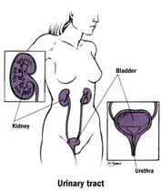
What is the urinary tract?

The "urinary tract" consists of the various organs of the body that produce, store, and get rid of urine. These include the kidneys, the ureters, the bladder, and the urethra.
Our kidneys are chemical filters for our blood. About one-quarter of the blood pumped by the heart goes through the kidneys. The kidneys filter this blood, and the "filtrate" is processed to separate out waste products and excess amounts of minerals, sugar, and other chemicals. Since it sees so much of the body's blood flow, the kidneys also contain pressure-sensitive tissue which helps the body control blood pressure, and some of the minerals and water are saved or discarded partly to keep your blood pressure in the proper range.
The waste products and "extras" make up the urine, which flows through "ureters" (one per kidney) into the bladder, where it is held until you are ready to get rid of it. When you urinate, muscles in the bladder wall help push urine out of the bladder, through the urethra, and out. (In men, the urethra passes through the penis; in women, the urethra opens just in front of the vagina.) When you aren't urinating (which is most of the time) a muscle called the "sphincter" squeezes the urethra shut to keep urine in; the sphincter relaxes when you urinate so that urine can flow out easily.
Urine is normally sterile -- that is, it does not normally contain bacteria. This is a good thing, since the mineral content of urine make it a great medium for bacteria to grow in. (If you have sugar in your urine, it's an even better culture medium, but that shouldn't happen unless you are diabetic, or are one of the rare people -- like me -- who are not diabetic but still have sugar in their urine.) Usually several things keep bacteria out of the urine. These include:
  • The urethral sphincter: when the urethra is squeezed shut, bacteria cannot climb up the urethra from the "meatus" (the outside opening) into the bladder.
  • The length of the urethra: it's a long way up to the bladder for a bacterium. (A woman's urethra is shorter than a man's, which is one reason why women are much more likely than men to get UTI's.)
  • Frequent washing: any bacteria that make it into the urethra are flushed out the next time you urinate, and since most people empty their bladders almost completely when they urinate any bacteria that get to the bladder will be flushed out too. There are also valves where the ureters enter the bladder to prevent urine from "refluxing" from the bladder to the kidneys, so even if the bladder and its urine is infected the bacteria shouldn't travel up to the kidneys

How does an infection start?

The urinary tract can be infected from above (by bacteria entering the kidneys from the bloodstream and travelling downward) or from below (by bacteria entering the urethra and travelling upward).
 
Infection from above is most often seen in newborns with generalized infection or sepsis. If there are many bacteria in the bloodstream, some are likely to get through the filters of the kidney to the urine. This is especially likely if the filters are immature, or if there are a lot of bacteria.
 
In older children and adults infection most often starts from below. In small children still using diapers, stool (which is largely bacteria) can sit for some time right at the meatus; the longer it sits there, the more likely it is that bacteria may enter the urethra. Baby boys are less likely to have this happen than baby girls, because girls' urethrae are much shorter and the head of the penis isn't as likely to sit in stool. (Note, though, that bacteria can hang out in any moist, warm area, and that UTI's in boys under 1 year old seem to happens more often in uncircumcised boys than in circumcised boys since bacteria can accumulate beneath the foreskin.) Older girls may become prone to UTI's through wiping back-to-front when they are first toilet-trained, which pulls stool into the vaginal/meatal area. Sexually active teenage and adult women are more prone to UTI's because of friction at the meatus, which tends to push bacteria into the urethra (urinating after intercourse helps avoid UTI's); the same mechanism may cause UTI's in teenage boys and adult men, although they are again less prone to UTI's than women of the same age.

Where do UTI's occur in the urinary tract?

In general, the farther the organ in the urinary tract from the place where the bacteria enter, the less likely the organ is to be infected.
  • Urethritis is infection/inflammation of the urethra. This can be due to other things besides the organisms usually involved in UTI's; in particular, many sexually-transmitted diseases (STD's) appear initially as urethritis. However, stool-related bacteria (the most common bacteria on the skin near the meatus) will also often cause urethritis.

  • Cystitis is an infection of the bladder. (Strictly speaking, "-itis" means inflammation, and there are non-bacterial reasons for bladder inflammation, but they are much less common than bacterial infection.) This is the most common form of UTI; it can be aggravated if the bladder does not empty completely when you urinate. (Some people have valves at the bladder end of the urethra as well as at the bladder ends of the ureters. You aren't supposed to have urethral valves except for the sphincter; these "extra" valves usually prevent complete bladder emptying and make cystitis more likely.)

  • Ureteritis is infection of a ureter. This can occur if the bacteria entered the urinary tract from above, or if the ureter-to-bladder valves don't work properly and allow urine to "reflux" from the bladder into the ureters.

  • Pyelonephritis is an infection of the kidney itself. This can happen with infection from above, or if reflux into the ureters is so bad that infected urine refluxes all the way to the kidney.

Symptoms of UTI's


The symptoms a person has with a UTI depend on how old the person is and on where in the urinary tract the infection is located.
Urethritis usually appears as burning on urination. Often this burning occurs mainly when you start urinating, since the bacteria and infected urine in the urethra cause the inflammation but are flushed out when "fresh" urine flows through the urethra on its way out of the bladder.
Cystitis may show up as burning on urination, often in the "middle" of urination. However, it may have no symptoms other than fever, lower abdominal (way down -- just above the pubic bone) pain, or even just a funny smell or colour or appearance (cloudy, dark, even blood-tinged) to your urine.
Blood in the urine can be a sign -- sometimes the only sign at first -- of a urinary tract infection. It can result from microscopic bleeding within the kidneys, or from an abscess if the infection is far advanced. Blood can also appear in urine from a bleed anywhere between the kidneys and the urinary meatus (the end of the urethra, from where the urine emerges); in particular, cystitis can result in bleeding inside the bladder, which will certainly leave blood in the urine -- whether as blood-tinging, blood clots in the urine, or something in between. When we ask patients what part of the urine stream the blood appears in, we are trying to figure out where the blood is entering the urine: for example, blood that appears just as you start to urinate and clears up as the flow continues indicates that the bleeding is in the urethra, where it accumulates until you urinate and is then flushed out by the flowing urine. On the other hand, blood that is uniformly mixed with the urine is likely coming from the kidneys, the ureters, or the bladder.
Since your kidneys are located in your back, just below your bottom ribs, pyelonephritis may appear as pain in your back or flank(s), or in the abdomen. Fever usually (but not always) comes along with the pain. If the kidneys are severely affected, you may also start seeing some of the complications due to kidney malfunction.


Complications of UTI's

Urinary tract infections can make you pretty miserable. They can do other things, too.
The biggest problem with a UTI is if it progresses to pyelonephritis. This can result in scarring and damage to the kidney tissue. Although the kidney's filter system is pretty big, it is not infinite. If there is enough damage to the filter system, waste products can't be removed properly. This constitutes kidney failure, and if it is bad enough and long-lasting enough the only solutions are dialysis (filtering your blood through an "artificial kidney" which isn't nearly as good as the real one and requires you to sit hooked up to a lot of plumbing three times a week) or a kidney transplant (which also poses many risks and problems).
A different complication occurs if the pressure-regulation tissues in the kidney are scarred. If this is bad enough, your blood pressure may be kept too low (and you'll faint frequently at the very least) or too high (leading to strokes, heart disease, and other nasty things).
Both of these problems may occur rapidly, but only if the infection is very severe. More often, the damage done by the initial infection, even if it is not compounded by future infections, progresses over many months or years. In particular, renal failure may not be complete until long after the first UTI

How do we treat (and evaluate) a UTI?

The first step in treating a UTI is to make sure there really is one. The only certain way to know if there is a UTI is to take a sample of urine and "culture" it: try to grow bacteria from the sample. If there are bacteria, we can then test several antibiotics to see which ones kill the bacteria most efficiently.
 
The problem here is in getting a good sample of urine for culture. Simply urinating into a sterile cup may not stop contamination by bacteria on the skin, especially with girls. If you can control your urine, it is possible to use a "clean-catch" sample. You get this by cleaning the meatus and the surrounding area thoroughly with antiseptics (such as iodine solution), then urinating a little into the toilet before filling the sample cup, and finishing your urination in the toilet. This flushes out bacteria that may be in the urethra or meatus.
 
Unfortunately, small children can't cooperate well enough to do this sort of collection, even if they are toilet-trained. We can collect urine with a bag ("puck") that is taped over the meatus and genitals. However, this almost guarantees contamination by skin bacteria. We sometimes use pucks to collect samples for follow-up culture, but such samples just don't work well for the initial diagnosis where we have to know whether or not there really is an infection. For the initial diagnosis in small children we usually use a sterile catheter inserted into the bladder through the urethra (after cleaning the meatal area with iodine or another soap that kills bacteria). This may sound barbaric, but it is the only way to be sure if a small child has a UTI or not. In newborn babies who may be septic, we may go even farther and draw urine out of the bladder with a needle inserted over the pubic bone (a suprapubic bladder tap) -- which may sound even more barbaric than the catheter, but the stakes are a lot higher in a newborn baby who doesn't have the defenses against infections that older children and adults have, and a suprapubic urine culture is postive if there are any bacteria growing in it -- no ifs, ands, or buts.
 
Once we have diagnosed a UTI we treat the patient with antibiotics. Typical antibiotics used for UTIs include trimethoprim-sulfamethoxamole, nitrofurantoin, ciprofloxacin, levofloxacin, or their chemical relatives, and certain penicillins such as amoxicillin. In some cases, when we are pretty sure from the symptoms that you actually have a UTI, we will start antibiotics right after we get the urine culture; if the culture result shows that we need a different antibiotic, we can always change. We often repeat the culture 3-5 days after starting antibiotics to make sure that we are actually killing all the bacteria, and again soon after the antibiotics are finished to make sure we killed everything that needed killing.
 
We also need to make sure that the infection did not get beyond the bladder, or, if it did, that the kidneys haven't been damaged. This is usually done with "nuclear scans" in which a tiny amount of a radioactive medicine is injected into the patient's bloodstream, where it heads for the kidneys to be excreted. The medicine can be detected with radiation detecting cameras, giving a picture of the kidneys: damaged kidney tissue will appear on the picture. (Older methods involving X-rays don't produce pictures nearly as good as the nuclear scan pictures, and expose you to much more radiation. The amount of radiation involved in nuclear kidney scans is much less than even standard X-rays would give.)
 
Ultrasound images of the kidneys, ureter, and bladder can show abscesses that may be present, as well as abnormalities in the "plumbing" (such as duplicate ureters or blocked ureters). It won't necessarily show the source of microscopic bleeding, but if the bleeding is microscopic it may stop after the infection is treated and we may never know precisely where the blood was entering the urine.

A voiding cystourethrogram, or "VCUG", is an X-ray of the kidneys and bladder taken after a "contrast medium" (a medicine which blocks X-rays) is injected into the bladder through a catheter in the urethra. We use the VCUG to look for reflux: if there is reflux, the contrast medium will go up into the ureters, and perhaps the kidneys it the reflux is severe, and this will be visible on the X-rays. As you can imagine, this isn't very comfortable for the patient, but the VCUG is the only practical way to find out if there is reflux. If reflux is bad enough, surgery can improve valve function and reduce reflux in some patients. Milder cases of reflux will often improve as a child grows; for intermediate grades of reflux we may decide to give a child low doses of antibiotics until the reflux improves (which may take several months). The antibiotics we use to treat UTIs are excreted from the body through the kidney and urine -- in fact, that's why we use those antibiotics -- so even low doses give levels of the antibiotic in the urine that are high enough to kill the few bacteria that might stray into the bladder, and resistance isn't as much of a problem as it might otherwise be.

Please Note-Cardiac patients who are on Anti Platelet drugs like Clopidogrel should consult with their doctor if they are suffering from URINARY TRACT INFECTION ESPECIALLY HAEMATURIA(appearance of blood in the urine).


HEART ATTACK


What Is a Heart Attack?

A heart attack is when blood vessels that supply blood to the heart are blocked, preventing enough oxygen from getting to the heart. The heart muscle dies or becomes permanently damaged. Your doctor calls this a myocardial infarction


A heart attack occurs when blood flow to a section of heart muscle becomes blocked. If the flow of blood isn’t restored quickly, the section of heart muscle becomes damaged from lack of oxygen and begins to die.
Heart attack is a leading killer of both men and women in the world. But fortunately, today there are excellent treatments for heart attack that can save lives and prevent disabilities. Treatment is most effective when started within 1 hour of the beginning of symptoms. If you think you or someone you’re with is having a heart attack, call emergency help  right away.

Overview


Heart attacks occur most often as a result of a condition called coronary artery disease (CAD). In CAD, a fatty material called plaque (plak) builds up over many years on the inside walls of the coronary arteries (the arteries that supply blood and oxygen to your heart). Eventually, an area of plaque can rupture, causing a blood clot to form on the surface of the plaque. If the clot becomes large enough, it can mostly or completely block the flow of oxygen-rich blood to the part of the heart muscle fed by the artery.

During a heart attack, if the blockage in the coronary artery isn’t treated quickly, the heart muscle will begin to die and be replaced by scar tissue. This heart damage may not be obvious, or it may cause severe or long-lasting problems.
Severe problems linked to heart attack can include heart failure and life-threatening arrhythmias (irregular heartbeats). Heart failure is a condition in which the heart can’t pump enough blood throughout the body. Ventricular fibrillation is a serious arrhythmia that can cause death if not treated quickly.

Get Help Quickly!!!!!

Acting fast at the first sign of heart attack symptoms can save your life and limit damage to your heart. Treatment is most effective when started within 1 hour of the beginning of symptoms

The most common heart attack signs and symptoms are:
  • Chest discomfort or pain—uncomfortable pressure, squeezing, fullness, or pain in the center of the chest that can be mild or strong. This discomfort or pain lasts more than a few minutes or goes away and comes back.
  • Upper body discomfort in one or both arms, the back, neck, jaw, or stomach.
  • Shortness of breath may occur with or before chest discomfort.
  • Other signs include nausea (feeling sick to your stomach), vomiting, lightheadedness or fainting, or breaking out in a cold sweat.
If you think you or someone you know may be having a heart attack:
  • Call Emergency within a few minutes—5 at the most—of the start of symptoms.
  • If your symptoms stop completely in less than 5 minutes, still call your doctor.
  • Only take an ambulance to the hospital. Going in a private car can delay treatment.
  • Take a nitroglycerin/ sorbitrate pill if your doctor has prescribed this type of medicine.

Treatment

If you had a heart attack, you will need to stay in the hospital, possibly in the intensive care unit (ICU). You will be hooked up to an ECG machine, so the health care team can look at how your heart is beating.
Life-threatening irregular heartbeats (arrhythmias) are the leading cause of death in the first few hours of a heart attack. These arrythmias may be treated with medications or electrical cardioverson/defibrillation.
The health care team will give you oxygen, even if your blood oxygen levels are normal. This is done so that your body tissues have easy access to oxygen and your heart doesn't have to work as hard.
An intravenous line (IV) will be placed into one of your veins. Medicines and fluids pass through this IV. You may need a tube inserted into your bladder (urinary catheter) so that doctors can see how much fluid your body removes.

ANGIOPLASTY AND STENT PLACEMENT

Angioplasty, also called percutaneous coronary intervention (PCI), is the preferred emergency procedure for opening the arteries for some types of heart attacks. It should preferably be performed within 90 minutes of arriving at the hospital and no later than 12 hours after a heart attack.
Angioplasty is a procedure to open narrowed or blocked blood vessels that supply blood to the heart.
A coronary artery stent is a small, metal mesh tube that opens up (expands) inside a coronary artery. A stent is often placed after angioplasty. It helps prevent the artery from closing up again. A drug eluting stent has medicine in it that helps prevent the artery from closing.

THROMBOLYTIC THERAPY (CLOT-BUSTING DRUGS)

Depending on the results of the ECG, certain patients may be given drugs to break up the clot. It is best if these drugs are given within 3 hours of when the patient first felt the chest pain. This is called thrombolytic therapy. The medicine is first given through an IV. Blood thinners taken by mouth may be prescribed later to prevent clots from forming.
Thrombolytic therapy is not appropriate for people who have:
  • Bleeding inside their head (intracranial hemorrhage)
  • Brain abnormalities such as tumors or blood vessel malformations
  • Stroke within the past 3 months (or possibly longer)
  • Head injury within the past 3 months
Thrombolytic therapy is extremely dangerous in women who are pregnant or in people who have:
  • A history of using blood thinners such as coumadin
  • Had major surgery or a major injury within the past 3 weeks
  • Had internal bleeding within the past 2-4 weeks
  • Peptic ulcer disease
  • Severe high blood pressure
OTHER MEDICINES FOR HEART ATTACKS

Many different medicines are used to treat and prevent heart attacks. Nitroglycerin helps reduce chest pain. You may also receive strong medicines to relieve pain.
Antiplatelet medicines help prevent clots from forming. Aspirin is an antiplatelet drug. Another one is clopidogrel (Plavix). Ask your doctor which of these drugs you should be taking. Always talk to your doctor before stopping either of these drugs.
  • For the first year after a heart attack, you will likely take both aspirin and clopidogrel every day. After that, your doctor may only prescribe aspirin.
  • If you had angioplasty and a coronary stent placed after your heart attack, you may need to take clopidogrel with your aspirin for longer than one year.
Other medications you may receive during or after a heart attack include:
  • Beta-blockers (such as metoprolol, atenolol, and propranolol) help reduce the strain on the heart and lower blood pressure.
  • ACE inhibitors (such as ramipril, lisinopril, enalapril, or captopril) are used to prevent heart failure and lower blood pressure.
  • Lipid-lowering medications, especially statins (such as lovastatin, pravastatin, simvastatin, atorvastatin, and rosuvastatin) reduce blood cholesterol levels to prevent plaque from increasing. They may reduce the risk of another heart attack or death.
Always talk to your doctor before stopping any medications, especially these drugs. Stopping or changing the amount of these medicines can be life threatening.

CORONARY ARTERY BYPASS SURGERY

Coronary angiography may reveal severe coronary artery disease in many vessels, or a narrowing of the left main coronary artery (the vessel supplying most of the blood to the heart). In these circumstances, the cardiologist may recommend emergency coronary artery bypass surgery (CABG). This procedure is also called "open heart surgery." The surgeon takes either a vein or artery from another location in your body and uses it to bypass the blocked coronary artery.

Outlook;

Each year, lakhs of people have heart attacks, and almost half of them die. CAD, which often results in a heart attack, is the leading killer of both men and women.
Many more people could recover from heart attacks if they got help faster. Of the people who die from heart attacks, about half die within an hour of the first symptoms and before they reach the hospital.


Common reasons for not seeking medical care
  • People believe only men have heart attacks.
    • Fact: Heart disease is the most common cause of death in women as well as men. If you have symptoms, call for help.
  • You are not sure it is a heart attack.
    • Call anyway--heart attack symptoms can be vague.
  • "I can't afford to go to the hospital"
    • Although hospital care is expensive, a heart attack effects your life and future health- this is priceless!
  • It feels like heartburn.
    • If you have a history of heart disease, angina, or high blood pressure, the heartburn you feel may actually be a heart attack.
      Call for help immediately.
  • You'd feel embarrassed if it turned out you didn't need medical help after all.
    • Never feel embarrassed about calling for help. A little embarrassment might save your life.
  • You're hoping that it is not a heart attack.
    • Wishful thinking can be deadly. You can't wish a heart attack away, and getting treatment quickly can save your life.
    • Most people who survive a heart attack can return to their normal life, including work and sexual activity, within 3 months.
  • I'm too young to have a heart attack
    • Although much less common in those under 40, heart attacks can strike at any age.
    • If you have any of the risk factors (especially stimulant drug use or family history) seek help for symptoms

PLEASE READ ARTICLES-'EMERGENCY CHEST PAIN' AND 'CARDIAC RESUSCITATION' FOR MORE INFORMATION .

MITRAL VALVE REPAIR AND REPLACEMENT SURGERY


Blood that flows between different chambers of your heart must flow through a valve. This valve is called the mitral valve. It opens up enough so blood can flow from one chamber of your heart (left atria) to the next chamber (left ventricle). It then closes, keeping blood from flowing backwards


The information provided below is about mitral valve replacement surgery resulting from mitral valve regurgitation, mitral valve prolapse, mitral valve stenosis and other heart valve disorders (including infection)
Mitral valve replacement is a complex surgical procedure designed to remove a diseased mitral valve from the heart. There are several different surgical approaches and options for patients to consider


Why do mitral valves need to be replaced?


Heart valves perform the important function of ensuring blood flow in the correct direction throughout the body. The mitral valve directs the flow of blood from the left atrium into the left ventricle, and the aortic valve allows blood to pass from the left ventricle into the aorta.
Mitral valve replacement typically results from two conditions known as stenosis and regurgitation. 


 Mitral valve stenosis


Mitral valve stenosis diagnosis suggests that the patient's valve fails to open properly due to stiff or rigid valve leaflets. Mitral stenosis can be caused from infection, calcification of the heart valve leaflets or tissue degeneration due to age.

Because a stenotic mitral valve may cause blood to back up into the lungs, careful monitoring of mitral valve stenosis symptoms should be monitored. Severely stenotic mitral valves may require mitral valve replacement surgery.

 Mitral valve regurgitation


Mitral Valve Repair and Replacement Surgery


Mitral valve replacement may also be required for patients suffering from mitral valve regurgitation also known as mitral insufficiency. When the mitral valve fails to close properly (primarily due to mitral valve prolapse), the need for mitral valve replacement is usually determined by (i) how severely the symptoms impact the patient and (ii) how well those symptoms can be controlled by medical treatment.
However, it should be noted that many patients who are asymptomatic may require mitral valve replacement surgery if their heart is dilated (enlarged) or experiencing other issues related to heart valve disease and/or congestive heart failure.

An operation to repair or replace a mitral valve takes 2-3 hours to perform. The damaged valve is either repaired and supported with a "ring" or it is replaced with either a "tissue" or "mechanical" valve . Although tissue and mechanical valves function similarly, there are distinct advantages and disadvantages of each. The advantage of mechanical valves, which are made from ceramic, is that they last forever. The disadvantage of mechanical valves is that they require anticoagulation with blood thinners for the remainder of a patient's life.
The advantage of tissue valves, which are made from cow or pig hearts, is that they do not require formal anticoagulation. The disadvantage of tissue valves, however, is that they generally wear out after 12-15 years, at which time another operation would be required to replace the worn out valve.


Procedure;Mitral Valve Repair/Replacement-open heart surgery


The operation itself requires general anesthesia ... the patient is asleep for the entire course of the operation. The surgeon opens the chest by dividing the breast bone or sternum. Tubes and cannulae are inserted into the heart and major blood vessels surrounding the heart in preparation for cardiopulmonary bypass with the heart-lung machine. At this point, blood is redirected from the heart into the heart-lung machine, the heart is stopped, and the aorta is clamped. This permits the surgeon to safely open and operate on the heart without blood pumping through it. The heart-lung machine continues to pump freshly oxygenated blood to the rest of the body, in effect, taking over the roles of the heart and lungs.
The surgeon then opens the heart, usually through the left atrium, and exposes the mitral valve. If the valve is only partially damaged, it is repaired and the rim or "annulus" of the valve is supported with a "ring". If, however, the valve cannot be repaired, it is removed. Non-absorbable sutures with bolsters or "pledgets" are used to sew the new valve into position . The valve is tested, to ensure that it opens and closes safely, and then the left atrium of the heart is closed. The clamp on the aorta is removed, and all air is evacuated from the heart. As the heart regains its strength, the patient is weaned from the heart-lung machine and the heart and lungs resume their normal functions


Procedure-Minimally invasive mitral valve surgery:

It is done through much smaller incisions (cuts) than the large incision needed for open surgery
Before your surgery you will receive general anesthesia. This will make you unconscious and unable to feel pain.
There are several different ways to perform minimally invasive mitral valve surgery.
  • Your heart surgeon may make a 2-inch to 3-inch-long incision (cut) in the right part of your chest near the sternum (breastbone). Muscles in the area will be divided so your surgeon can reach the heart. A small cut is made in the left side of your heart so the surgeon can repair or replace the mitral valve.
  • In endoscopic surgery, your surgeon makes 1 to 4 small holes in your chest. Then your surgeon uses special instruments and a camera to do the surgery.
  • For robotically-assisted valve surgery, the surgeon makes 2 to 4 tiny cuts (about ½ to ¾ inch) in your chest. The surgeon uses a special computer to control robotic arms during the surgery. The surgeon sees a 3-dimensional view of the heart and aortic valve on the computer. This method is very precise.
You will not need to be on a heart-lung machine for these types of surgery, but your heart rate will be slowed by medicine or a mechanical device.
If your surgeon can repair your aortic valve, you may have:
  • Ring annuloplasty -- The surgeon repairs the ring-like part around the valve by sewing a ring of metal, cloth, or tissue around the valve.
  • Valve repair -- The surgeon trims, shapes, or rebuilds 1 or more of the 3 leaflets of the valve. The leaflets are flaps that open and close the valve.


If your mitral valve is too damaged, you will need a new valve. This is called replacement surgery. Your surgeon will remove your mitral valve and sew a new one into place. There are two main types of new valves:
  • Mechanical -- made of man-made materials, such as cloth, metal, or ceramic. These valves last the longest, but you will need to take blood-thinning medicine, such as warfarin (Coumadin) or aspirin, for the rest of your life.

  • Biological -- made of human or animal tissue. These valves last 12 to 15 years, but you may not need to take blood thinners for life.

The surgery may take 1 to 3 hours.
This surgery can also be done through a groin artery, with no incisions on your chest. The doctor sends a catheter (flexible tube) with a balloon attached on the end. The balloon stretches the opening of the valve. This procedure is called percutaneous valvuloplasty.

Why the Procedure is Performed:


You may need surgery if your mitral valve does not work properly.
  • A mitral valve that does not close all the way will allow blood to leak back into the left atria. This is called mitral regurgitation.
  • A mitral valve that does not open fully will restrict blood flow. This is called mitral stenosis.
Minimally invasive surgery may be done for these reasons:
  • Changes in your mitral valve are causing major heart symptoms, such as angina (chest pain), shortness of breath, syncope (fainting spells), or heart failure.
  • Tests show that the changes in your mitral valve are beginning to seriously affect your heart function.
  • Your heart valve has been damaged by endocarditis (infection of the heart valve).
A minimally invasive procedure has many benefits. There is less pain, blood loss, and risk of infection. You will also recover faster than you would from open heart surgery.


Risks:


Risks for any surgery are:
  • Reactions to medicines
  • Breathing problems
  • Blood clots in the legs that may travel to the lungs
  • Infection, including in the lungs, kidneys, bladder, chest, or heart valves
  • Blood loss
Minimally invasive surgery techniques have far fewer risks than open surgery. Possible risks from minimally invasive valve surgery are:
  • Irregular heartbeat that must be treated with medicines or a pacemaker
  • Damage to other organs, nerves, or bones
  • Heart attack, stroke, or death


Before the Procedure;


Always tell your doctor or nurse:
  • If you are or could be pregnant
  • What drugs you are taking, even drugs, supplements, or herbs you bought without a prescription
You may be able to store blood in the blood bank for transfusions during and after your surgery. Ask your surgeon about how you and your family members can donate blood.
For the 2-week period before surgery, you may be asked to stop taking drugs that make it harder for your blood to clot. These might cause increased bleeding during the surgery.
  • Some of them are aspirin, ibuprofen (Advil, Motrin), and naproxen (Aleve, Naprosyn).
  • If you are taking warfarin (Coumadin) or clopidogrel (Plavix), talk with your surgeon before stopping or changing how you take these drugs.
Prepare your house for when you get home from the hospital.
The day before your surgery. Shower and shampoo well. You may be asked to wash your whole body below your neck with a special soap. Scrub your chest 2 or 3 times with this soap. You also may be asked to take an antibiotic, to guard against infection.

During the days before your surgery:
  • Ask your doctor which drugs you should still take on the day of your surgery.
  • If you smoke, you must stop. Ask your doctor for help.
  • Always let your doctor know if you have a cold, flu, fever, herpes breakout, or any other illness in the time leading up to your surgery.
On the day of the surgery:
  • You will usually be asked not to drink or eat anything after midnight the night before your surgery. This includes chewing gum and using breath mints. Rinse your mouth with water if it feels dry, but be careful not to swallow.
  • Take the drugs your doctor told you to take with a small sip of water.
  • Your doctor or nurse will tell you when to arrive at the hospital.


After the Procedure


Expect to spend 3 to 5 days in the hospital after surgery. You will wake up in the intensive care unit (ICU) and recover there for 1 or 2 days. Nurses will closely watch monitors that show information about your vital signs (pulse, temperature, and breathing).
Two to 3 tubes will be in your chest to drain fluid from around your heart. They are usually removed 1 to 3 days after surgery. You may have a catheter (flexible tube) in your bladder to drain urine. You may also have intravenous (IV, in a vein) lines to get fluids.
You will go from the ICU to a regular hospital room. Your nurses and doctors will continue to monitor your heart and vital signs until you are stable enough to go home. You will receive pain medicine for pain in your chest.
Your nurse will help you slowly resume some activity. You may begin a program to make your heart and body stronger.
A temporary pacemaker may be placed in your heart if your heart rate becomes too slow after surgery

Outlook (Prognosis)


Mechanical heart valves do not fail often. However, blood clots develop on them. If a blood clot forms, you may have a stroke. Bleeding can occur, but this is rare.
Biological valves tend to fail over time. But they have a lower risk of blood clots.
Techniques for minimally invasive heart valve surgery have improved greatly over the past 10 years. These techniques are safe for most patients, and they reduce recovery time and pain.


AORTIC VALVE-REPLACEMENT AND REPAIR SURGERY

AORTIC VALVE SURGERY

Blood flows out of your heart and into the aorta through a valve. This valve is called the aortic valve. It opens up so blood can flow out. It then closes, keeping blood from flowing backwards.

Aortic valve surgery is done to either repair or replace the aortic valve in your heart.
  • An aortic valve that does not close all the way allows blood to leak back into your heart. This is called aortic regurgitation.
  • An aortic valve that does not open fully will restrict blood flow. This is called aortic stenosis.
Minimally invasive aortic valve surgery is done through much smaller incisions (cuts) than the large cut needed for open aortic valve surgery.


Minimally invasive aortic valve surgery


Description;


Before your surgery you will receive general anesthesia. This will make you unconscious and unable feel pain.
There are several different ways to do minimally invasive aortic valve surgery. Techniques include laparoscopy or endoscopy, robot-assisted surgery, and percutaneous surgery.
  • Your surgeon may make a 2-inch to 3-inch-long incision (cut) in the right part of your chest near the sternum (breastbone). Muscles in the area will be divided to so your surgeon can reach the valve. This allows the surgeon to reach your heart and aortic valve.
  • For the endoscopic, or “keyhole, approach, your surgeon makes 1 to 4 small holes in your chest. Then your surgeon uses special instruments and a camera to do the surgery.
  • For robotically-assisted valve surgery, the surgeon makes 2 to 4 tiny cuts (about ½ to ¾ inch) in your chest. The surgeon uses a special computer to control robotic arms during the surgery. The surgeon sees a 3-dimensional view of the heart and aortic valve on the computer. This method is very precise.
You will not need to be on a heart-lung machine for any of these surgeries, but your heart rate will be slowed by medicine or a mechanical device.
If your surgeon can repair your aortic valve, you may have:
  • Ring annuloplasty -- The surgeon repairs the ring-like part around the valve by sewing a ring of metal, cloth, or tissue around the valve.


  • Valve repair -- The surgeon trims, shapes, or rebuilds 1 or more of the 3 leaflets of the valve. The leaflets are flaps that open and close the valve.

If your aortic valve is too damaged, you will need a new valve. This is called replacement surgery. Your surgeon will remove your aortic valve and sew a new one into place. There are two main types of new valves:
  • Mechanical -- made of man-made materials, such as cloth, metal, or ceramic. These valves last the longest, but you will need to take blood-thinning medicine, such as warfarin (Coumadin) or aspirin, for the rest of your life.


  • Biological -- made of human or animal tissue. These valves last 12 to 15 years, but you may not need to take blood thinners for life.

Once the new or repaired valve is working, your surgeon will
  • Close the small cut to your heart or aorta
  • Place catheters (flexible tubes) around your heart to drain fluids that build up
  • Close the surgical cut in your muscles and skin
The surgery may take 1 to 3 hours.
Aortic valve surgery is also now being done through a groin artery. No incisions are made on your chest. The doctor sends a catheter (tube) with a balloon attached on the end to the valve. The balloon stretches the opening of the valve. This procedure is called percutaneous valvuloplasty.

A minimally invasive procedure has many benefits. There is less pain, blood loss, and risk of infection. You will also recover faster than you would from open heart surgery.

Techniques for minimally invasive heart valve surgery have improved greatly over the past 10 years.

Open Aortic Valve Surgery;


Description:


In open surgery, the surgeon makes a large incision (cut) in your breastbone to reach the heart and aorta
Before your surgery you will receive general anesthesia. This will make you unconscious and unable to feel pain.
  • Your surgeon will make a 10-inch-long cut in the middle of your chest.
  • Next, your surgeon will separate your breastbone to be able to see your heart and aorta (the main blood vessel leading from your heart to the rest of your body).
  • Most people are connected to a heart-lung bypass machine or bypass pump. Your heart is stopped while you are connected to this machine. This machine does the work of your heart while your heart is stopped.
If your surgeon can repair your aortic valve, you may have:
  • Ring annuloplasty -- The surgeon repairs the ring-like part around the valve by sewing a ring of metal, cloth, or tissue around the valve.
  • Valve repair -- The surgeon trims, shapes, or rebuilds 1 or more of the 3 leaflets of the valve. The leaflets are flaps that open and close the valve.
If your aortic valve is too damaged, you will need a new valve. This is called replacement surgery. Your surgeon will remove your aortic valve and sew a new one into place. There are two main types of new valves:
  • Mechanical -- made of man-made materials, such as cloth, metal, or ceramic. These valves last the longest, but you will need to take blood-thinning medicine, such as warfarin (Coumadin) or aspirin, for the rest of your life.
  • Biological -- made of human or animal tissue. These valves last 12 to 15 years, but you may not need to take blood thinners for life.

Once the new or repaired valve is working, your surgeon will
  • Close your heart and take you off the heart-lung machine.
  • Place catheters (tubes) around your heart to drain fluids that build up.
  • Close your breastbone with stainless steel wires. It will take about 6 weeks for the bone to heal. The wires will stay inside your body.
This surgery may take 3 to 6 hours.
Sometimes other procedures are done during open aortic about surgery. These include the Ross (or switch) procedure, the David procedure, and a graft of the ascending aorta

Why the Procedure is Performed;


You may need surgery if your aortic valve does not work properly. You may need open-heart valve surgery for these reasons:
  • Changes in your aortic valve are causing major heart symptoms, such as angina (chest pain), shortness of breath, syncope (fainting spells), or heart failure.
  • Tests show that changes in your aortic valve are beginning to seriously harm how well your heart works.
  • Your heart valve has been damaged by endocarditis (infection of the heart valve).
  • You have received a new heart valve in the past and it is not working well, or you have other problems such as blood clots, infection, or bleeding.


Risks:

Risks for any anesthesia are:
  • Reactions to medicines
  • Breathing problems
  • Blood clots in the legs that may travel to the lungs
  • Infection, including in the lungs, kidneys, bladder, chest, or heart valves
  • Blood loss
Possible risks from having open heart surgery and minimally invasive aortic valve surgery are similar;
  • Heart attack or stroke
  • Incision infection, which is more likely to occur in people who are obese, have diabetes, or have already had this surgery
  • Post-pericardiotomy syndrome, which is a low-grade fever and chest pain. This could last up to 6 months.
  • Memory loss and loss of mental clarity, or "fuzzy thinking."
  • Heart rhythm problems


Before the Procedure:


Always tell your doctor or nurse:
  • If you are or could be pregnant
  • What drugs you are taking, even drugs, supplements, or herbs you bought without a prescription
You may be able to store blood in the blood bank for transfusions during and after your surgery. Ask your surgeon about how you and your family members can donate blood.
For the 2-week period before surgery, you may be asked to stop taking drugs that make it harder for your blood to clot. These might cause increased bleeding during the surgery.
  • Some of them are aspirin, ibuprofen (Advil, Motrin), and naproxen (Aleve, Naprosyn).
  • If you are taking warfarin (Coumadin) or clopidogrel (Plavix), talk with your surgeon before stopping or changing how you take these drugs.
During the days before your surgery:
  • Ask your doctor which drugs you should still take on the day of your surgery.
  • If you smoke, you must stop. Ask your doctor for help.
  • Always let your doctor know if you have a cold, flu, fever, herpes breakout, or any other illness in the time leading up to your surgery.
Prepare your house for when you get home from the hospital.
The day before your surgery, shower and shampoo well. You may be asked to wash your whole body below your neck with a special soap. Scrub your chest 2 or 3 times with this soap. You also may be asked to take an antibiotic, to prevent infection.

On the day of your surgery:
  • You will usually be asked not to drink or eat anything after midnight the night before your surgery. This includes chewing gum and using breath mints. Rinse your mouth with water if it feels dry, but be careful not to swallow.
  • Take the drugs your doctor told you to take with a small sip of water.
  • Your doctor or nurse will tell you when to arrive at the hospital.

After the Procedure:

Expect to spend 5 to 7 days in the hospital after surgery. You will wake up in the intensive care unit (ICU) and stay there for 1 or 2 days. Two to 3 tubes will be in your chest to drain fluid from around your heart. These are usually removed 1 to 3 days after surgery.
You may have a catheter (flexible tube) in your bladder to drain urine. You may also have intravenous (IV, in a vein) lines to deliver fluids. Nurses will closely watch monitors that show information about your vital signs (your pulse, temperature, and breathing).
You will be moved to a regular hospital room from the ICU. Your nurses and doctors will continue to monitor your heart and vital signs until you are stable enough to go home. You will receive pain medicine to control pain around your incision.
Your nurse will help you slowly resume some activity. You may begin a program to make your heart and body stronger.
A temporary pacemaker may be placed in your heart if your heart rate becomes too slow after surgery.

Outlook(Prognosis)

Mechanical heart valves do not fail often. However, blood clots develop on them. If a blood clot forms, you may have a stroke. Bleeding can occur, but this is rare.

Biological valves tend to fail over time. But they have a lower risk of blood clots