Cochin Cardiac Club

Health Blog by Dr.Uday Nair

MUSCULAR DYSTROPHY (MD)


Muscular dystrophy (MD) is a group of more than 30 inherited diseases. They all cause muscle weakness and muscle loss. Some forms of MD appear in infancy or childhood. Others may not appear until middle age or later. The different types can vary in whom they affect, which muscles they affect, and what the symptoms are. All forms of MD grow worse as the person's muscles get weaker. Most people with MD eventually lose the ability to walk.

There is no cure for muscular dystrophy. Treatments can help with the symptoms and prevent complications. They include physical and speech therapy, orthopedic devices, surgery, and medications. Some people with MD have mild cases that worsen slowly. Others cases are disabling and severe.


Types of muscular dystrophy


There are many different types of MD, each with somewhat different symptoms. Not all types of MD cause severe disability and many do not affect life expectancy.

Some of the more common types of MD include:

  • Duchenne muscular dystrophy – one of the most common and severe forms, it usually affects boys in early childhood; men with the condition will usually only live into their 20s or 30s
  • Myotonic dystrophy – a type of MD that can develop at any age; life expectancy is not always affected, but people with a severe form of it may have shortened lives
  • Facioscapulohumeral muscular dystrophy – a type of MD that can develop in childhood or adulthood, it progresses slowly and is not usually life threatening
  • Becker muscular dystrophy – closely related to Duchenne MD, but it develops later in childhood and is less severe; life expectancy is not usually affected so much
  • Limb-girdle muscular dystrophy – a group of conditions that usually develop in late childhood or early adulthood; some variants can progress quickly and be life threatening, whereas others only develop slowly
  • Oculopharyngeal muscular dystrophy – a type of MD that usually doesn't develop until a person is 50-60 years old and doesn't tend to affect life expectancy
  • Emery-Dreifuss muscular dystrophy – a type of MD that develops in childhood or early adulthood; most people with this condition will live until at least middle age


Symptoms of Muscular Dystrophy


The symptoms of muscular dystrophy are the result of a deterioration of the body's muscles. This deterioration is due to the death of the muscle cells and muscle tissues and leads to ongoing muscle wasting and muscle weakness. Muscular dystrophy progresses and gets worse over time eventually. This results in difficulty walking, disability, the need for leg and hand braces, and ultimately the use of a wheelchair. 

The muscle weakness of muscular dystrophy often begins in the legs. This makes it difficult for a child to walk normally, and he or she may walk with their feet wide apart to help keep balance. The child may use his or her hands and arms to get up from the floor and assist with standing. There may be frequent falls, a waddling gait, limited range of motion and pain in the calves. By 12 years of age, a child is often completely unable to walk and must use a wheelchair. 


Diagnosis


The diagnosis of muscular dystrophy is based on the results of muscle biopsy, increased creatine phosphokinase (CpK3), electromyography, electrocardiography and DNA analysis.
A physical examination and the patient's medical history will help the doctor determine the type of muscular dystrophy. Specific muscle groups are affected by different types of muscular dystrophy.
Often, there is a loss of muscle mass (wasting), which may be hard to see because some types of muscular dystrophy cause a build up of fat and connective tissue that makes the muscle appear larger. This is called pseudohypertrophy.

Treatment


There's currently no cure for any form of muscular dystrophy. Research into gene therapy may eventually provide treatment to stop the progression of some types of muscular dystrophy. Current treatment is designed to help prevent or reduce deformities in the joints and the spine and to allow people with muscular dystrophy to remain mobile as long as possible.
Medications
Corticosteroids, such as prednisone, may help improve muscle strength and delay the progression of certain types of muscular dystrophy. But prolonged use of these types of drugs can weaken bones and increase fracture risk.
Therapy
Several different types of therapy and assistive devices can improve quality and sometimes length of life in people who have muscular dystrophy. Examples include:
Surgical remedies are an option for several of the problems common to muscular dystrophy, such as:
  • Contractures. Tendon surgery can loosen joints drawn inward by contractures.
  • Scoliosis. Surgery may also be needed to correct a sideways curvature of the spine that can make breathing more difficult.
  • Heart problems. Some people who have heart problems related to muscular dystrophy may be helped by the insertion of a pacemaker, which prompts the heart to beat more regularly.
  • Range-of-motion exercises. Muscular dystrophy can restrict the flexibility and mobility of joints. Limbs often draw inward and become fixed in that position. One goal of physical therapy is to provide regular range-of-motion exercises to keep joints as flexible as possible.
  • Mobility aids. Braces can provide support for weakened muscles and help keep muscles and tendons stretched and flexible, slowing the progression of contractures. Other devices — such as canes, walkers and wheelchairs — can helfp maintain mobility and independence.
  • Breathing assistance. As respiratory muscles weaken, a sleep apnea device may help improve oxygen delivery during the night. Some people with severe muscular dystrophy may need to rely on a ventilator — a machine that forces air in and out of their lungs.

Potential Treatment for Muscular Dystrophy Patients Through Stem Cell Therapy


While there is no cure to date for muscular dystrophy, stem cell research and implantation of healthy embryonic cells into individuals diagnosed with various forms of muscular dystrophy may improve function and mobility as well as slow the muscle-wasting process.

There are a number of different types of stem cells that scientists think may be used in different ways to develop treatments for muscular dystrophy. The main stem-cell-based approaches currently being investigated are:
  1. Producing healthy muscle fibres: Scientists hope that if stem cells without the genetic defect that causes DMD can be delivered to patients’ muscles, they may generate working muscle fibres to replace the patient’s damaged ones.
  2. Reducing inflammationIn muscular dystrophy damaged muscles become very inflamed. This inflammation speeds up muscle degeneration. Scientists believe certain types of stem cells may release chemicals that reduce inflammation, slowing the progress of the disease.
Beside stem cells, other therapeutic strategies such as gene therapy or small-molecule drugs for repairing the damaged gene are being tested in patients and in pre-clinical models. Future therapies are likely to use a combination of more than one of these approaches. Scientists are also studying the role of stem cells in the maintenance and repair of healthy muscles in order to understand in more detail what goes wrong in muscular dystrophy and how the problem could be corrected.
Research, clinical trials, therapies and treatments in stem cell implantation and transplants will continue in this field as researchers around the globe search for greater understanding of the best type of stem cells to be utilized in treating all forms of muscular dystrophy, offering hope to millions affected with the muscle wasting disease.


CHEST PAIN AFTER ANGIOPLASTY AND CORONARY ARTERY BYPASS GRAFTING(CABG)


Coronary Angioplasty:

A coronary angioplasty is also referred to as PCI or percutaneous coronary intervention. It is performed in order to open up heart arteries that are clogged. This procedure may be accompanied by the placement of a stent to help keep the artery open. A stent is a small metal coil that may or may not be treated with a coating of medication to aid in the function of the stent.

Chest pain after angioplasty with stent is a common occurrence. It can simply be part of the healing process or something more serious.

Causes of Chest pain after Angiolasty


  • Ischemia Following Angioplasty with Stent

    Ischemia is the condition in which the blood supply become restricted, usually due to blood vessels being blocked or having some other problem that inhibits blood flow.
    When ischemia occurs after angioplasty, it may be called “silent ischemia”. One of the ways ischemia is treated is with angioplasty with the placement of a stent. When ischemia follows angioplasty with stent placement, the procedure may be performed again.
  • Restenosis Following Angioplasty with Stent

    Restenosis is the name given to the condition in which the artery treated with angioplasty begins to close. This can happen even when a stent is in place. Restenosis can cause chest pain in the patient when the patient exerts himself or herself.
    The treatment for restenosis is usually a repeat surgery.

    Management of Chest pain after Angiolasty


  • Chest pain following successful percutaneous coronary interventions is a common problem. Although the development of chest pain after coronary interventions may be of benign character, it is disturbing to patients, relatives and hospital staff. Such pain may be indicative of acute coronary artery closure, coronary artery spasm or myocardial infarction, but may also simply reflect local coronary artery trauma. The distinction between these causes of chest pain is crucial in selecting optimal care.Management of these patients may involve repeat coronary angiography and additional intervention. Commonly, repeat coronary angiography following percutaneous transluminal coronary angioplasty (PTCA) in patients with chest pain demonstrates widely patent lesion sites suggesting that the pain was due to coronary artery spasm, coronary arterial wall stretching or was of non-cardiac origin.

  • Please Note


    If the chest pain increases or is at the site of the procedure, the doctor should be contacted immediately.
  • If chest pain is experienced after angioplasty with stent placement, the treating physician should be contacted in order to determine whether it is due to recovery or something else.


    Coronary Artey Bypass Grafting(CABG):

    Coronary bypass surgery is a procedure that restores blood flow to your heart muscle by diverting the flow of blood around a section of a blocked artery in your heart. Coronary bypass surgery uses a healthy blood vessel taken from your leg, arm, chest or abdomen and connects it to the other arteries in your heart so that blood is bypassed around the diseased or blocked area. After a coronary bypass surgery, blood flow to your heart is improved. 

    Coronary artery bypass graft surgery (CABG) is performed in patients with stable angina and acute coronary syndromes to prolong life or to treat angina refractory to medical or percutaneous revascularization therapies. Angina may return after apparently successful CABG. 

    Causes of Chest Pain after CABG


    The cause varies with the time when symptoms are first noted after surgery: 

  • Recurrent angina during the early postoperative period is usually due to a technical problem with a graft or with early graft closure. This is indication for prompt coronary angiography with percutaneous coronary intervention (PCI), if feasible.
  • Recurrent angina after the first few months, called late recurrent angina, can occur with the development of stenosis in a bypass graft or with progression of atherosclerosis in non-bypassed vessels.

  • Management


    Chest wall pain after cardiac surgery may normally last 3-6 weeks, but may last as long as 12 weeks on rare occasions.This pain has a different quality from angina and most patients know the difference without question.  Post-op pain is also related to movement, coughing, and normally can be reproduced with palpation of the chest wall, none of which occur with ischemic pain (angina).It’s extremely rare to have angina following CABG (coronary artery bypass grafting), and differentiating between angina and post-op pain should be simple with just a physical exam.

    Please Note


    The discomfort you feel following coronary bypass surgery is typically related to motion and coughing.Don’t let chest pain, that follows coronary bypass surgery, alarm you, even if you’re experiencing it weeks after. However, it’s important to note concerning discomfort to your surgeon and cardiologist.


    TRANSCATHETER AORTIC VALVE REPLACEMENT(TAVR)


    Transcatheter Aortic Valve Replacement (TAVR), also known as Transcatheter Aortic Valve Implantation (TAVI), is an innovative approach to replacing the aortic valve without open-heart surgery.This is a revolutionary new treatment option for patients with severe aortic stenosis who are not well enough to undergo traditional open heart surgery. The TAVR procedure gives hope for extending the lives of these patients with improved quality of life.This heart valve replacement is minimally invasive which means a majority of patients will experience faster recovery times than traditional open-heart valve replacement.


    Aortic Stenosis


    Patients who suffer from aortic stenosis- a progressive disease that affects the aortic valve of the heart- often experience symptoms that can restrict normal day-to-day activities, such as walking short distances or climbing stairs. Severe aortic stenosis is a serious problem. Patients who do not receive an aortic valve replacement (AVR) have no effective, long-term treatment option to prevent or delay their disease progression. Without it, severe aortic stenosis is life threatening – studies indicate that 50 percent of patients will not survive more than an average of two years after the onset of symptoms.


    TAVR Criteria


    Until now, the only treatment for severe aortic stenosis has been open aortic valve replacement, but if a cardiac surgeon determines that you are not a candidate for open-heart surgery and if medicine is not helping you feel better, the TAVR procedure may be an alternative. TAVR is approved for patients who have shortness of breath or chest pain from a severely narrowed aortic valve and who are to sick for an open aortic valve operation.Most people who have this procedure are in their 70s or 80s and often have other medical conditions that make them a better candidate for this type of surgery. 


    TAVR Procedure


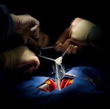
    During the valve replacement procedure, a cardiologist inserts a tube (catheter) through an artery in the groin (transfemoral approach) or a small incision between the ribs (transapical approach).The transcatheter heart valve is compressed into the end of the catheter.The valve is made of bovine or cow tissue and polyester supported with a stainless-steel mesh frame. Once released from the catheter, the heart valve is expanded with a balloon and immediately becomes functional.The valve is designed to replace a patient's diseased native aortic valve without traditional open-heart surgery and while the heart continues to beat – eliminating the need for cardiopulmonary bypass. 


    Potential Benefits


    • Fragile patients can tolerate this procedure
    • Less time spent in surgery
    • Less pain and quicker recovery time
    • Lower risk of complications


    Risks


    TAVR is a significant procedure typically requiring general anesthesia. While less invasive than open heart surgical aortic valve replacement, TAVR still has the potential for similar adverse effects, including risk of death, stroke, damage to the artery used for replacement of the valve, major bleeding, and other life-threatening and serious events. The components of the TAVR valve are made from similar material as those in a conventional aortic valve replacement.


    TUBERCULOSIS - ( TB )


    Tuberculosis (TB), a multisystemic disease with myriad presentations and manifestations, is the most common cause of infectious disease–related mortality worldwide. Although TB rates are decreasing, the disease is becoming more common in many parts of the world. In addition, the prevalence of drug-resistant TB is increasing worldwide.

    Causes


    Tuberculosis (TB) is caused by a type of bacterium called mycobacterium tuberculosis.
    TB is spread when a person with an active TB infection in their lungs coughs or sneezes and someone else inhales the expelled droplets containing TB bacteria.
    However, although it is spread in a similar way to cold or flu, TB is not as contagious. You would usually have to spend prolonged periods in close contact with an infected person to catch the infection yourself.
    For example, TB infections usually spread between family members who live in the same house. It would be highly unlikely to become infected by sitting next to an infected person on a bus or train.
    Not everyone with TB is infectious. Generally, children with TB or people with TB that occurs outside the lungs (extrapulmonary TB) do not spread the infection.

    Latent or active TB

    Your immune system will usually be able to defeat the bacteria that cause TB. However, in some cases the bacteria infect the body but don't cause any symptoms (latent TB) or the infection will begin to cause symptoms within weeks or months (active TB).
    About 10% of people with latent TB develop active TB years after the initial infection. This usually happens when the immune system is weakened, for example during chemotherapy.

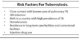
    Risk Factors


    Anyone can catch TB, but people particularly at risk include:
    • those living in environments where the level of existing TB infection is higher than normal
    • people with health conditions such as HIV or whose circumstances mean they are less able to fight off a TB infection
    Other things that can increase your risk of developing an active TB infection include:
    • being in close contact with someone who is infected
    • having lived in, travelled to or had visitors from parts of the world where TB is common
    • being part of an ethnic group that originated in parts of the world where TB is still common
    • having a weakened immune system because of HIV, diabetes or other medical conditions
    • having a weakened immune system because of long courses of medication, such as corticosteroids, chemotherapy or tumour necrosis factor blockers (used to treat some types of arthritis) 
    • being very young or very old – the immune systems of people who are young or elderly tend to be weaker than those of healthy adults
    • being in poor health or having a poor diet due to lifestyle and other problems, such as drug misuse, alcohol misuse or homelessness
    • living in poor or crowded housing conditions, such as prisons

    Signs and Symptoms of TB


    People with a latent TB infection don’t have any symptoms, don’t feel sick and cannot infect others. They do, however, test positive to the Mantoux Skin Test. Treating latent TB is important since it can get activated, especially if the immune system is weakened for any reason, including nutritional deficiencies or infection with HIV.
    In the case of an active infection, signs and symptoms vary according to the organ that is affected.
    In case the lungs are affected, the symptoms are:
    • A cough persisting for 2 to 3 weeks and beyond, which is usually worse in the mornings
    • Chest pain
    • Blood in the sputum (the mucus and saliva produced when coughing or clearing throat)
    • Breathlessness
    Back pain may be caused by tuberculosis of the spine, and blood in the urine may be caused by tuberculosis in your kidneys.
    TB in the brain can cause headaches, a stiff neck, confusion, vomiting, an altered mental state, seizures and other signs and symptoms related to the nerves.
    In general, a person with active TB in any organ may have these signs and symptoms:
    • Weight loss
    • Loss of appetite
    • Chills
    • Fever
    • Sweating in the night time while sleeping, even if the weather is cold.
    Diagnosis


    Tuberculosis is diagnosed by finding Mycobacterium tuberculosis bacteria in a clinical specimen taken from the patient. While other investigations may strongly suggest tuberculosis as the diagnosis, they cannot confirm it.
    A complete medical evaluation for tuberculosis (TB) must include a medical history, a physical examination, a chest X-ray and microbiological examination (of sputum or some other appropriate sample). It may also include a tuberculin skin test, other scans and X-rays, surgical biopsy.

    Treatment


    Medications are the cornerstone of tuberculosis treatment. But treating TB takes much longer than treating other types of bacterial infections. With tuberculosis, you must take antibiotics for at least six to nine months. The exact drugs and length of treatment depend on your age, overall health, possible drug resistance, the form of TB (latent or active) and the infection's location in the body.
    A recent study suggests that a shorter term of treatment — three months instead of nine — with combined medication may be effective in keeping latent TB from becoming active TB. With the shorter course of treatment, people are more likely to take all their medication and the risk of side effects is lessened. More study is needed.
    Most common TB drugs
    If you have latent tuberculosis, you may need to take just one type of TB drug. Active tuberculosis, particularly if it's a drug-resistant strain, will require several drugs at once. The most common medications used to treat tuberculosis include:
    • Isoniazid
    • Rifampin (Rifadin, Rimactane)
    • Ethambutol (Myambutol)
    • Pyrazinamide
    There's some evidence that taking vitamin D during tuberculosis treatment enhances some of the effects of the drugs. More study is needed.
    Medication side effects
    Side effects of TB drugs aren't common but can be serious when they do occur. All tuberculosis medications can be highly toxic to your liver. When taking these medications, call your doctor immediately if you experience any of the following:
    • Nausea or vomiting
    • Loss of appetite
    • A yellow color to your skin (jaundice)
    • Dark urine
    • A fever that lasts three or more days and has no obvious cause
    Completing treatment is essential
    After a few weeks, you won't be contagious, and you may start to feel better. It might be tempting to stop taking your TB drugs. But it is crucial that you finish the full course of therapy and take the medications exactly as prescribed by your doctor. Stopping treatment too soon or skipping doses can allow the bacteria that are still alive to become resistant to those drugs, leading to TB that is much more dangerous and difficult to treat.
    To help people stick with their treatment, a program called directly observed therapy (DOT) is sometimes recommended. In this approach, a health care worker administers your medication so that you don't have to remember to take it on your own.
    Prevention

    Tuberculosis is a preventable and a curable disease if detected and treated early.Some measures of prevention are suggested for those living or travelling to areas and countries that have a high prevalence of the infection.

    Tuberculosis vaccine

    Bacillus Calmette-Guérin (BCG) vaccination can protect against tuberculosis. The BCG vaccine is given to all infants in countries where the disease is prevalent. In countries where it is less common it is given to those who are at risk.
    Before the vaccine is given, the person is given a Mantoux skin test to check for latent tuberculosis. Vaccination is not recommended for people with latent tuberculosis.

    Currently, the BCG vaccination is recommended for three main groups of people. One of these are babies born in areas where the rates of tuberculosis is high and those babies with one or more parents or grandparents born in countries with a high rate of tuberculosis. Mantoux skin test will not be required beforehand while giving BCG vaccine to a baby.
    Another group that needs BCG vaccine includes children under 16 years of age who have one or more parents or grandparents born in countries with a high rate of tuberculosis and have not been vaccinated as babies. Children under 16 who have been in close contact with someone with tuberculosis or have lived for at least three months in a country with a high rate of tuberculosis are also vaccinated with BCG after getting a Mantoux test.
    The third group that requires BCG vaccination are those at high-risk occupations that includes people under 35 years of age whose occupation expose them to tuberculosis infected persons. This includes:-
    • health care workers (doctors, nurses and carers)
    • laboratory personnel who handle samples of tuberculosis
    • veterinary staff and other animal workers, such as abattoir workers who handle animals that may carry tuberculosis
    • those working in closed cramped spaces including prisoners, and hostel wardens, staff at orphanages and homes for the homeless etc.
    • staff at elderly care homes and refugee homes
    • travellers to countries with high rates of tuberculosis who plan to live there for at least 3 months

    Prevention of transmission of infection

    Those with pulmonary tuberculosis are contagious up to about two to three weeks once their treatment is begun. Earlier these patients were isolated. These days isolation is not practiced but some precautions are important to prevent spread. These include:-
    • Isolation from workplaces, schools and college and areas with crowds.
    • Covering one’s mouth and nose while coughing or sneezing.
    • Adequate and careful disposal of tissues. Usually burning or disposal in sealed plastic bags is recommended.
    • Sharing beds and rooms with un-infected persons while sleeping should be avoided.

    Please Note


    Early detection and treatment of TB is hugely important to stop the spread of this highly contagious disease.Make sure you know the symptoms and risks, so if TB finds a way into your home or workplace, you know what to do.About 1 in 10 latent cases, which present no symptoms, develop into full-blown TB. Knowing who is at risk can be almost as important as knowing what to look for.
    If your workplace or environment puts you into one of these high-risk groups, a TB test can be a smart way to prevent developing the disease.
    If you, or someone you know, does develop symptoms, it’s important to know what to look for. Most, but not all, infections are located mostly in the lungs, and lead to chestpain,difficulty breathing, and most noticeably, prolonged coughing (often with blood).  Those with TB may also experience flu-like symptoms, including chills, aches, fatigue, weight loss, and pallor.
    Drug-resistance is rapidly becoming a challenge in the treatment of TB, so quelling outbreaks early is extremely important. Early detection is one of the best steps toward stopping the spread of this preventable disease. Know what to look for and educate those around you, so everyone can breath easy.